| Cat # | Size | Price | Quantity | |
|---|---|---|---|---|
| 120803 | 25 tests | ¥1,290 | ||
| 120804 | 100 tests | ¥2,915 |
| Clone | 24F.10C12 |
|---|---|
| Application | Flow Cytometry |
| Reactivity | Human |
| Format | PE |
| Target Name | CD273, B7-DC, PD-L2, PDL2, B7DC |
| Isotype | Mouse IgG2a |
| Antibody Type | Monoclonal |
| Regulatory Status | RUO |
| Formulation | Phosphate-buffered solution, pH 7.2, containing 0.09% sodium azide and 0.2% (w/v) BSA |
| Protein Concentration | Supplied at a lot-specific concentration. |
| Storage & Handling | The antibody solution should be stored undiluted between 2°C and 8°C, and protected from prolonged exposure to light. Do not freeze. |
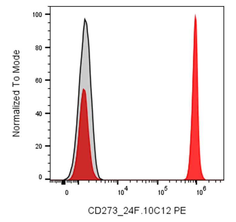
| Recommended Usage | For flow cytometric staining, it is recommended to use 5 µL of this reagent per 0.5-1.0 million cells in a 100 µL volume. Optimal reagent performance should be determined by titration for each specific application. PE has an excitation max at 565 nm and an emission max at 575 nm. |
| Excitation Laser | Blue Laser (488 nm) Green/Yellow laser (532/561nm) |
| See All Formats | Clone 24F.10C12 |
Human programmed death-ligand 2 (PD-L2), also known as CD273 or B7-DC, is an immune checkpoint protein encoded by the PDCD1LG2 gene. It belongs to the B7 family of immunoregulatory molecules and plays an important role in regulating T-cell responses and maintaining immune balance. PD-L2 is expressed primarily by antigen-presenting cells, including dendritic cells and macrophages, and can also be induced on certain other cell types under inflammatory conditions. By interacting with its receptor, PD-L2 can modulate T-cell activation, cytokine production, and immune tolerance.
PD-L2 is a type I transmembrane glycoprotein composed of an extracellular region containing two immunoglobulin-like domains, a single transmembrane segment, and a short intracellular cytoplasmic tail. Its principal established receptor is programmed cell death protein 1 (PD-1, CD279), expressed on activated T cells and other immune cells. Binding of PD-L2 to PD-1 delivers inhibitory signals that reduce T-cell receptor signaling, cytokine production, and proliferation, thereby limiting excessive immune activation. PD-L2 has a higher binding affinity for PD-1 than its related ligand PD-L1 in many experimental systems. Additional interactions, including binding to RGMb (repulsive guidance molecule b), have been reported and may contribute to immune regulation in particular tissues.
Dysregulated PD-L2 expression can influence disease progression. In cancer, some tumors and tumor-associated immune cells express PD-L2, enabling suppression of antitumor T-cell responses and potentially contributing to immune evasion. PD-L2 expression has been reported in several malignancies, although its prevalence and clinical significance vary by tumor type. In inflammatory and autoimmune conditions, PD-L2-mediated signaling may help restrain damaging immune responses, while altered regulation can affect immune tolerance and inflammation.
PD-L2 is a potential target for cancer immunotherapy. Blocking PD-L2, alone or alongside PD-1/PD-L1 pathway inhibitors, may restore antitumor T-cell activity in selected settings. Researchers are also investigating PD-L2 expression as a biomarker for immune activity and treatment response. Human PD-L2 is widely used in immunology and oncology research to study immune checkpoint signaling, T-cell regulation, and therapeutic strategies targeting the tumor immune microenvironment.
PE Mouse IgG2a Isotype Control Antibody
PE Anti-human CD273 (B7-DC, PD-L2) Antibody TDS
Have a product or application question? Consult our FAQs or contact us.